
Figure 1.
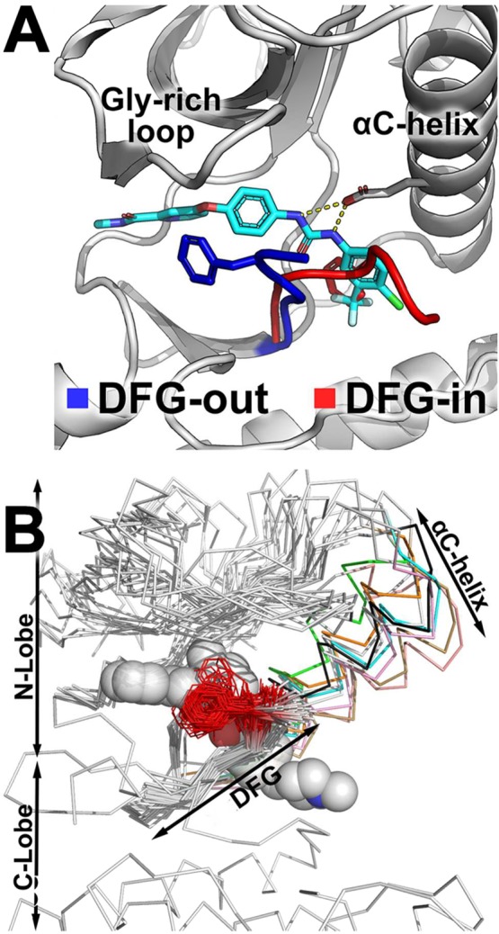
DFG-motif flips between two conformations. (A) Type-II inhibitor sorafenib (cyan sticks) is not compatible with the DFG-in conformation because it overlaps with the phenylalanine residue of the DFG-motif (red stick). (B) The DFG-motif and the residues preceding it are structurally conserved across most kinases, with the Phe side chain (red line) pointing away from the protein core. Type-II inhibitors (gray spheres) occupy the ATP-binding site and the DFG-pocket. The αC-helix of the N-lobe (colored ribbons) can adopt a wide range of conformations, thereby shifting the position of the N-lobe (white ribbons) relative to the C-lobe.