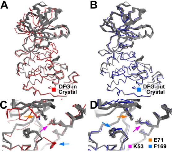
Figure 4.

DFGmodel models of P38α. Fifty models (gray ribbon) are shown superposed onto the C-lobe of a (A) DFG-in crystal structure (PDB: 2LGC) and a (B) DFG-out crystal structure (PDB: 4A9Y). (C, D) DFG-flip moves F169 (blue arrow) in the models by 11 Å. Centroid of the models’ N-lobe is shifted, displacing the conserved K53 (orange arrow) by 3.0 Å and E71 (violet arrow) on αC-helix by 1.0 Å. The DFGmodel models are structurally more similar to known DFG-out structures than to DFG-in structures.
