
FIG 2.
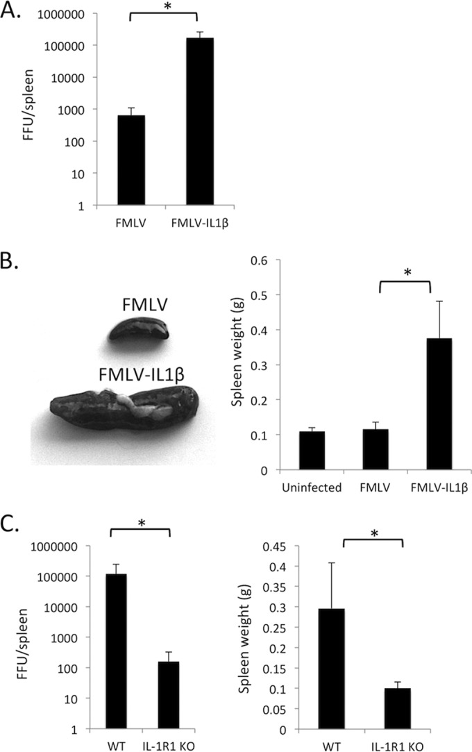
FMLV-IL-1β exhibits enhanced replication in vivo. (A) C57BL/6 (B6) mice were infected with FMLV or FMLV-IL-1β. At 7 dpi, mice were dissected, and the number of focus-forming units (FFU) per spleen was measured by plating splenocyte dilutions on Mus dunni cells followed by a focus-forming assay. The results of the focus-forming assay were used to calculate FFU/spleen in each infected mouse. (B) The weights of spleens from FMLV- and FMLV-IL-1β-infected mice were measured (right). Representative spleens are shown on the left. (C) Wild-type B6 (WT) or congenic IL-1 receptor 1-deficient (IL-1R1 KO) mice were infected with FMLV-IL-1β. At 7 dpi, FFU per spleen (left) and spleen weights (right) were measured by plating splenocyte dilutions on Mus dunni cells followed by a focus-forming assay. Each bar represents the average for 5 mice. The asterisks indicate P values of <0.05 (t test or Mann-Whitney test). Error bars indicate the standard deviations of the data set.