FIG 4.
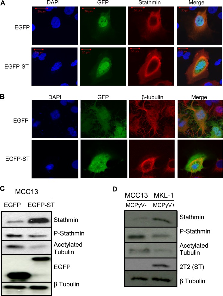
MCPyV ST promotes microtubule destabilization. MCC13 cells were transfected with either EGFP or EGFP-ST expression vectors. After 24 h, cells were fixed and permeabilized, and GFP fluorescence was analyzed by direct visualization, whereas endogenous stathmin (A) and endogenous β-tubulin (B) were identified by indirect immunofluorescence using stathmin- and β-tubulin-specific antibodies, respectively. (C) MCC13 cell lysates expressing EGFP or EGFP-ST were analyzed by immunoblotting using stathmin-, phosphorylated stathmin-, acetylated tubulin-, GFP-, and β-tubulin-specific antibodies. (D) Cellular lysates from MCC13 (MCPyV-negative) and MKL-1 (MCPyV-positive) cells were analyzed by immunoblotting using stathmin-, phosphorylated stathmin-, acetylated tubulin-, 2T2-, and β-tubulin-specific antibodies.

