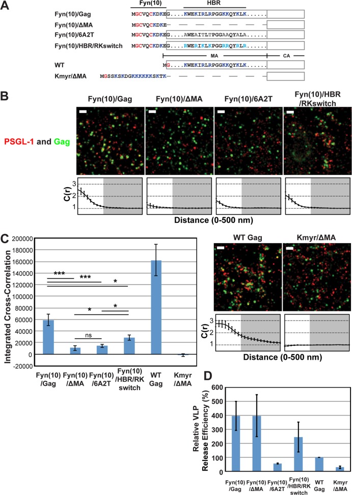
FIG 2.
Coclustering of Gag and PSGL-1 in HeLa cells is dependent on the intact MA HBR sequence but does not correlate with Gag membrane binding or VLP production. (A) Schematic representation of the MA mutants examined in this study. The amino acid residues of the Fyn(10), HBR, and Kmyr sequences are shown. Blue, basic residues; gray, uncharged amino acids; turquoise, switched residues; red, lipid-modified residues. (B) HeLa cells were transfected with plasmids carrying the indicated Gag-mEos3.2 fusion constructs and wild-type PSGL-1. Cells were fixed and immunostained for PSGL-1 with a monoclonal antibody against PSGL-1, which was directly labeled with Alexa Fluor 647, and imaged by TIRF microscopy in a reducing buffer (7,500 image frames per cell). Representative reconstructed images were produced as described in Materials and Methods. Images show Gag-mEos3.2 in green and PSGL-1 in red. Bars = 500 nm. Cross-correlation curves are shown below their corresponding representative images. Cross-correlation measurements were performed as described in the legend to Fig. 1A using images of a total of at least 10 cells per condition from 2 independent experiments. (C) Total coclustering was calculated as described in the legend to Fig. 1B. Values shown indicate means ± SEMs. ***, P < 0.0005; *, P < 0.05; ns, not significant. (D) A VLP release assay was performed on Gag-mEos3.2 constructs in HeLa cells. Results represent the averages of three independent experiments normalized to the value for WT Gag-mEos3.2.

