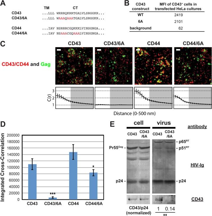
FIG 4.
Coclustering of CD43 and CD44 with HIV-1 Gag is enhanced by juxtamembrane polybasic sequences. (A) Partial amino acid sequences of CD43 and CD44 mutants. (B) The mean fluorescence intensities of HeLa cells expressing transfected CD43 constructs were determined as described in the legend to Fig. 3C. Note that the mean fluorescence intensities of HeLa cells transfected with CD44 expression plasmids were not determined due to a high endogenous expression of CD44. (C) HeLa cells were transfected with plasmids carrying WT Gag-mEos3.2 and the indicated constructs and imaged as described in the legend to Fig. 2. Representative reconstructed images and cross-correlation curves are shown. Bars = 500 nm. Gag-mEos3.2 is shown in green, and the indicated uropod-directed proteins or their derivatives are shown in red. (D) The analysis of total coclustering was performed as described in the legend to Fig. 1B using cross-correlation curves for images of a total of 10 cells per condition from 2 independent experiments. Values shown indicate means ± SEMs. P values were calculated for each mutant construct relative to its wild-type counterpart. ***, P < 0.0005; *, P < 0.05. (E) HeLa cells were transfected with a WT HIV-1 molecular clone (pNL4-3) and expression plasmids carrying wild-type CD43 or CD43/6A and cultured for 16 h. Cell and virus lysates were collected and subjected to immunoblotting analysis using HIV-Ig and anti-CD43. Note that no obvious difference in virus release efficiency was observed between CD43- and CD43/6A-expressing cells. The results shown are representative of the results from three independent experiments. The normalized CD43/p24 ratio shown at the bottom was calculated and averaged from the results of the three experiments. **, P < 0.005. RT, reverse transcriptase.

