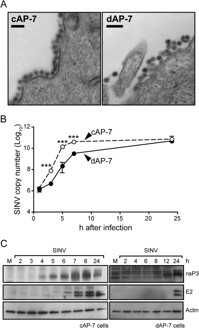
FIG 2.
SINV intracellular replication is delayed in differentiated AP-7 neurons. AP-7 neurons were infected with the TE strain of SINV (MOI, 5). (A) cAP-7 and dAP-7 neurons were fixed at 24 h after infection, and ultrastructure analysis was performed by transmission electron microscopy of thin sections. Bar, 100 nM. (B) Total cellular RNA was collected at various times after infection. cDNA was produced using SINV-specific primers and quantified by qPCR compared to standard SINV genomic DNA. SINV RNA levels in cAP-7 (dashed lines) or dAP-7 (solid lines) neurons are expressed as the mean SINV copy number (log10) ± standard deviation of triplicate samples from a representative of two experiments. (C) Immunoblot analysis of total cell lysates prepared at the indicated times from mock-infected (M) or SINV-infected cAP-7 (left) or dAP-7 (right) neurons with anti-nsP3 (top), anti-E2 (middle), or anti-β-actin (bottom) antibodies. A representative of 2 experiments is shown. Significant differences between cAP-7 and dAP-7 at each time point were determined by two-way ANOVA with Bonferroni's posttest. ***, P < 0.001.

