FIG 7.
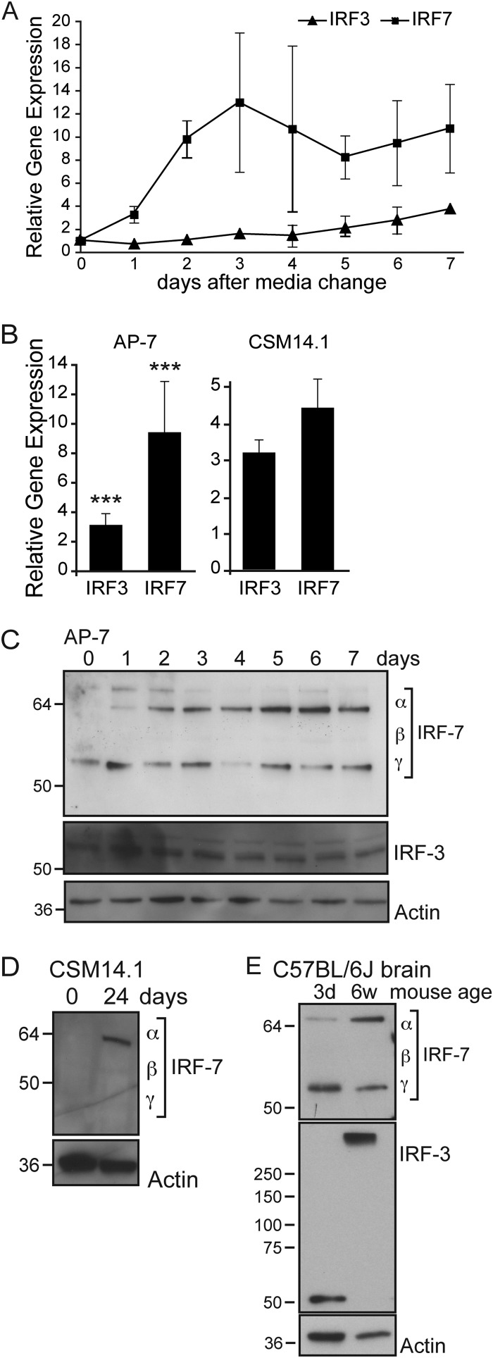
Irf-3 and Irf-7 mRNAs and IRF-7 protein increase during differentiation. (A) Total cellular RNA was collected at various times after plating of AP-7 neurons. Neurons were incubated at the permissive temperature in DMEM–10% FBS, 1 day prior to day 0 collection, corresponding to cAP-7 neurons. After day 0, neurons were maintained in differentiation medium at the nonpermissive temperature as described for the differentiation procedures. cDNA was produced using random primers, and Irf-3 and Irf-7 mRNAs were measured by qPCR and normalized to glyceraldehyde 3-phosphate dehydrogenase levels. mRNA levels are expressed as the mean value compared to levels in uninfected day 0 cAP-7 neurons ± standard deviations of six samples. (B) Total RNA was collected from day 0 (9 replicates) and day 7 (12 replicates) AP-7 neuron cultures or from triplicate cultures of day 0 or day 28 CSM14.1 neurons. mRNA levels, measured as described for panel A, are expressed as the mean value of differentiated cultures compared to levels in day 0 AP-7 or CSM14.1 neurons ± standard deviations. (C) Total cell lysates prepared from AP-7 neurons during differentiation were analyzed by immunoblot analysis with anti-IRF-7 (top row), anti-IRF-3 (middle row), or anti-β-actin (bottom row) antibodies. Multiple isoforms of IRF-7 are detected by the IRF-7 antibody. (D) Total cell lysates prepared from CSM neurons at the indicated days during differentiation were analyzed by immunoblot analysis with anti-IRF-7 (top row) or anti-β-actin (bottom row) antibodies. (E) Brain homogenates (10%) from 3-day-old or 6-week-old C57BL/6J mice were analyzed by immunoblot analysis with anti-IRF-7 (top row), anti-IRF-3 (middle row), or anti-β-actin (bottom row) antibodies. Results of a representative experiment of at least 2 experiments are shown. Significant differences between cAP-7 and dAP-7 at each time point were determined by one-way ANOVA with Dunnett's posttest. ***, P < 0.001.

