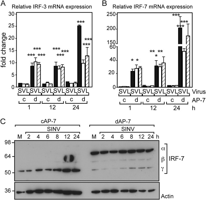
FIG 9.
Expression of Irf-3 and Irf-7 is upregulated during infection of differentiated AP-7 neurons. cAP-7 or dAP-7 neurons were infected with SINV (MOI, 5), VEEV (MOI, 50), or LACV (MOI, 5). Total cellular RNA was collected at 1, 12, or 24 h after infection, and cDNA was produced using random primers. Irf-3 (A) or Irf-7 (B) gene expression was measured by qPCR and normalized to glyceraldehyde-3-phosphate dehydrogenase levels. RNA levels are expressed as the mean value compared to levels in mock-infected cAP-7 neurons ± standard deviation of triplicate samples. Results of a representative experiment of 2 experiments are shown. Significant differences by two-way ANOVA with Bonferroni's posttest are shown. *, P < 0.05; **, P < 0.01; ***, P < 0.001. (C) cAP-7 or dAP-7 neurons were mock infected or infected with SINV (MOI, 5). Total cell lysates were prepared at the indicated times after infection and analyzed by immunoblotting using anti-IRF-7 (top) or anti-β-actin (bottom) antibodies. Results of a representative experiment of 2 experiments are shown.

