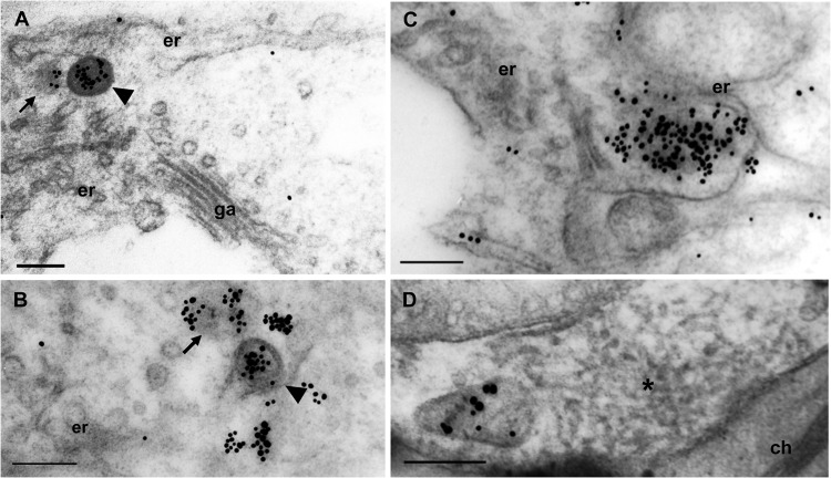
FIG 2.
NP forming cytoplasmic agglomerates in close proximity to the ER and NP agglomerates predominantly enveloped by the ER membrane in FMV-infected fig leaves. Immunogold labeling was performed using an anti-NP antibody and 15-nm gold particle-conjugated anti-rabbit antibody. (A and B) A DMB (arrowhead) and an agglomerate that is not enveloped (arrowhead). (C) Larger agglomerates curving the ER cisternae. (D) NP was not detected in the matrix (asterisk). er, endoplasmic reticulum; ch, chlorophyll. Bars, 200 nm.

