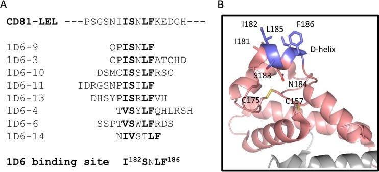
FIG 6.
Mapping of the 1D6 binding site to the D-helix of CD81. (A) Peptide sequences isolated from phage display screening with 1D6. Residues in bold represent the consensus located on CD81. (B) Cartoon representation of CD81-LEL structure (31). Residues I182, S183, L185, and F186, which compose the 1D6 binding site as presented in panel A, are highlighted, with carbons in metallic blue, oxygens in red, and nitrogens in blue. Other residues of CD81-LEL are also presented, with carbons in pale pink, oxygens in red, nitrogens in blue, and sulfurs in yellow. Conserved disulfide bond pairs are shown between cysteine residues at positions 175-157 and 156-190. The figure was generated using PyMOL.

