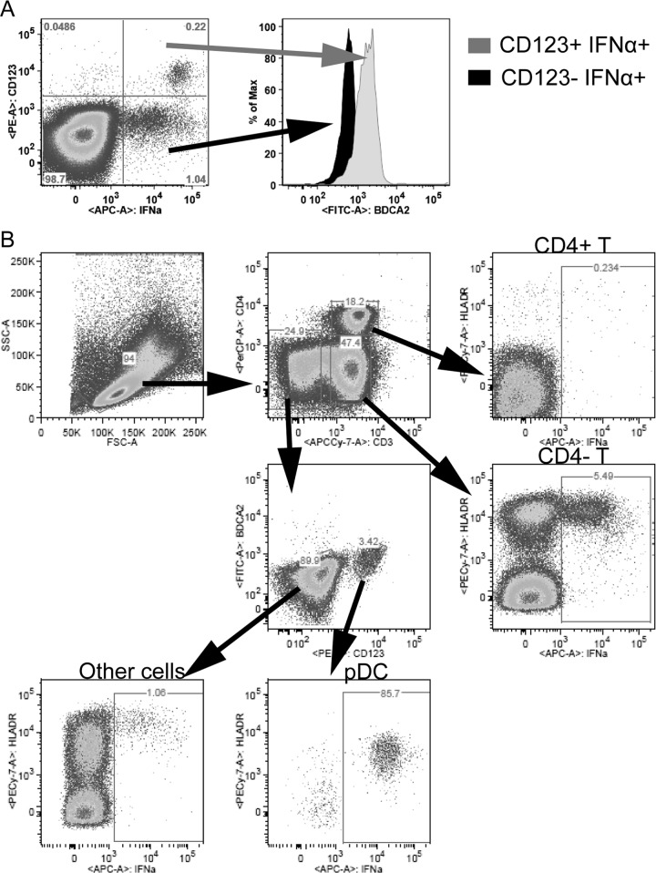
FIG 4.
(A) Identification of cell types producing IFN-I upon HSV stimulation. (A) BDCA-2 expression on IFN-α2+ cells. PBMCs from a representative animal stimulated with HSV at day 2 p.i. are shown. From the CD123+ IFN-α+ (gray) and the CD123− IFN-α+ (black) quadrants, the expression of BDCA-2 is shown. (B) PBMCs were gated for distinct cell populations to identify cell types that produced IFN-α2. PBMCs from one AGM at day 2 p.i. are shown. A total of 5.5% of the CD4− T cells, 85.7% of the pDCs, and 1% of non-T, non-pDCs were IFN-α+ after HSV stimulation. All IFN-α-producing cells expressed high levels of HLA-DR. The identified cells producing IFN-α in this animal and at this time point are representative of data from days 2, 9, and 11 postinfection.

