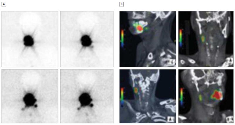
Figure 2. Comparison of 2 Imaging Methods.

Dynamic planar lymphoscintigraphy (A) and fused single-photon emission computed tomography/computed tomography (B) images from participant 5 demonstrating the delineation of sentinel lymph node location in relationship to adjacent structures in the 2 imaging modalities.
