
Figure 2.
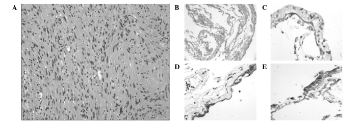
Photomicrographs of the solid mass illustrating (A) a benign spindle cell neoplasm with palisading of bland, vesicular nuclei consistent with a schwannoma (hematoxylin and eosin stain; original magnification, ×200); (B) the cyst wall, consisting of glial cells lined by a simple cuboidal to columnar epithelium (hematoxylin and eosin stain; original magnification, ×100); and positive staining for (C) cytokeratin, (D) glial fibrillary acidic protein and (E) S-100 protein (original magnification, ×200).