
Figure 3.
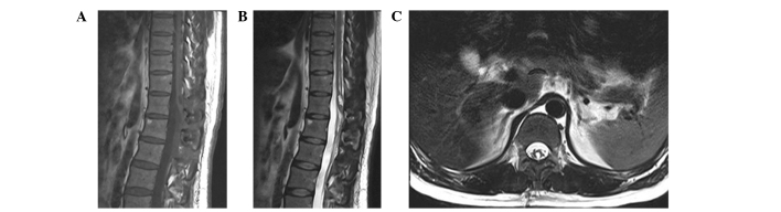
Contrast-enhanced (A) T1-weighted, (B) T2-weighted and (C) coronary T2-weighted magentic resonance images at nine months postsurgery showing no recurrence of the schwannoma or the cyst, and indicating the cavity left by the removal of the mass in the spinal cord.