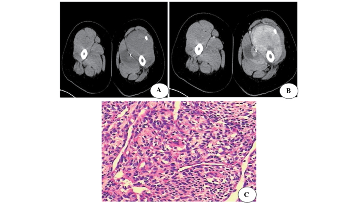
Figure 1.
A 31-year-old male with synovial sarcoma. (A) Computed tomography revealing a lobulated tumor mass with a low-intensity signal in the muscle of the left upper thigh. The tumor is well-defined and has punctate calcification. (B) Contrast-enhanced scan revealing heterogeneous enhancement and non-enhancement in areas of necrosis. (C) Pathological confirmation of synovial sarcoma carried out by hematoxylin and eosin staining.

