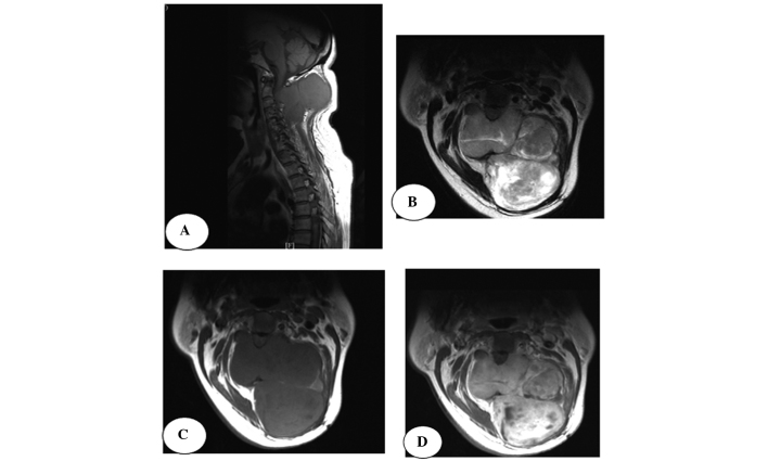
Figure 3.
A 56-year-old female with synovial sarcoma. (A) Magnetic resonance imaging revealing a well-defined lobulated soft-tissue mass in the neck. (B and C) T1-weighted imaging (T1WI) and T2-WI revealing a slightly hyperintensive signal relative to muscle and a hypointensitive signal indicating internal septations. (D) Contrast-enhanced scan revealing heterogeneous enhancement, while no clear enhancement is observed in the areas of necrosis and internal septations. Osseous destruction is located in adjacent vertebrae.

