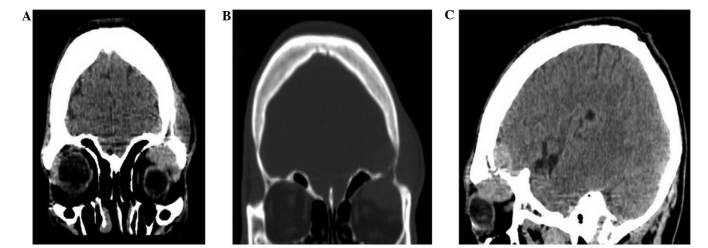
Figure 1.

Computed tomography imaging of the patient, including: (A) A coronal plane image demonstrating intracranial and extracranial masses in the left orbit region under the scalp; (B) a coronal plane image indicating lytic changes in the skull bone; and (C) a sagittal plane image showing a cranio-orbital mass.
