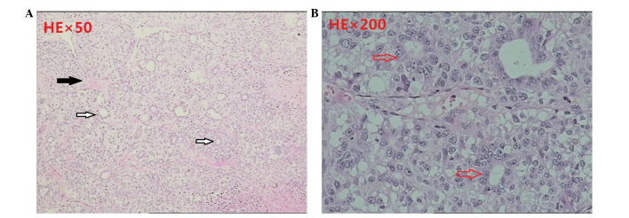
Figure 5.

Postoperative pathology of the excised metastatic lesion, showing (A) the portal area (black arrow), bile ducts (white arrow; magnification, ×50); and (B) nest-shaped cell clusters with central lumen that adopted the shape of the bile ducts (arrow; magnification, ×200; hematoxylin and eosin staining).
