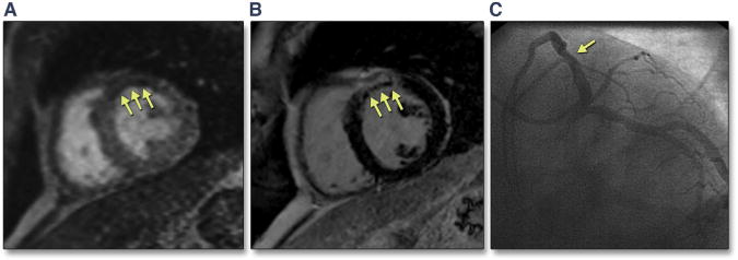
FIGURE 1. Cardiac Magnetic Resonance (CMR) Images From a 46-Year-Old Man With Diabetes and Chest Pain.

(A) First-pass perfusion (FPP) image shows a region of hypoperfusion in the anteroseptum (early microvascular obstruction [EMVO]). (B) Phase-sensitive inversion recovery (PSIR) sequence reveals presence of myocardial infarction (MI) with large area of late microvascular obstruction (LMVO). These findings were consistent with an acute MI in a diagonal branch that was originally missed on (C) cardiac catheterization (arrow shows proximal diagonal branch obstruction).
