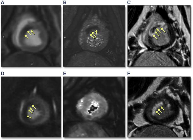
FIGURE 2. Example Images From a Study of Reperfused Acute MI in a Porcine Model.

The images from the first animal (top) show (A) decreased perfusion in the mid-anteroseptal wall on FPP imaging, (B) an area of intramyocardial hemorrhage on T2* mapping, and (C) a corresponding hypointense area on phase-sensitive late gadolinium enhancement imaging, consistent with microvascular obstruction (MVO) with IMH. Images from the second animal (bottom) (D) demonstrate an area of reduced perfusion on FPP imaging, (E) T2* maps do not demonstrate any IMH, and (F) late gadolinium enhancement images demonstrate the absence of MVO or IMH.
