
FIGURE 4. Images From a Mongrel Dog in Which MI Was Experimentally Induced.
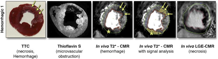
CMR was performed day 3 after reperfusion in which T2*-weighted gradient echo imaging was performed. Ex vivo, thioflavin S imaging, and triphenyl tetrazolium chloride staining were performed to assess for MVO, hemorrhage, and myocardial necrosis. Adapted with permission from Kumar et al. (45). CMR = cardiac magnetic resonance; LGE = late gadolinium enhancement; TTC = triphenyl tetrazolium chloride.