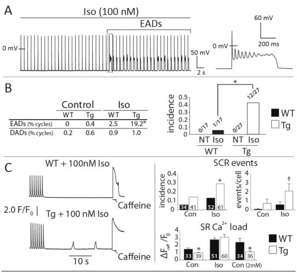
Figure 1.
CaMKIIδC myocytes are susceptible to EADs and SCR. A. Representative Tg cell transitioning to EADs during Iso challenge. B. EAD frequency (% of all cycles, left) and incidence (right) was increased in Tg compared to WT cells during Iso. Neither group exhibited EADs in Normal Tyrode’s (NT). C. Left: representative Ca2+ recordings during pause-induced SR Ca2+ release experiments. Right: summary data for SCR incidence (fraction of cells with 1 or more SCR events), SCR frequency (events/cell), and SR Ca2+ content at baseline, during Iso, and in elevated extracellular Ca2+ (2 mM, SR Ca2+ content only). Cell numbers are inset to each bar. **p < 0.01, *p < 0.05, † p = 0.06. Data are mean±SEM.

