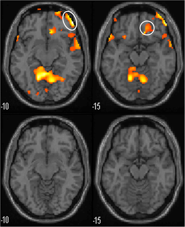
Figure 7.

Functional magnetic resonance imaging in activated PFC and ACC in moxibustion group during 100 ml rectal balloon distention. Upper row are the pictures before treatment, lower row are the pictures after treatment. The prefrontal cortex(PFC) and anterior cingulated cortex(ACC) are encircled. Left row is PFC and right row is ACC.
