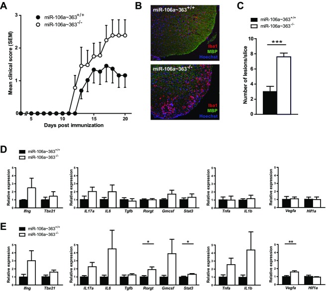
Figure 5.
EAE in miR-106a∼363-deficient mice. (A) Active EAE was induced in miR-106a∼363-deficient C57BL/6 mice using MOG35–55 peptide (n = 6–7). (B) Representative images of CNS lesions with mononuclear cell infiltration and microglia/macrophage activation. (C) Quantification of number of lesions per slice (n = 5). (D) Quantitative PCR from splenocytes extracted from animals at day 20 post immunization. The different panels represent targets associated to (from left to right) Th1 and Th17/GMCSF differentiation, as well as the macrophage lineage, all previously validated targets of miR-20b. (E) Quantitative PCR from spinal cord tissue at day 20 post immunization. Statistical tests were performed using t-test. EAE, experimental autoimmune encephalomyelitis; CNS, central nervous system.

