Figure 6.
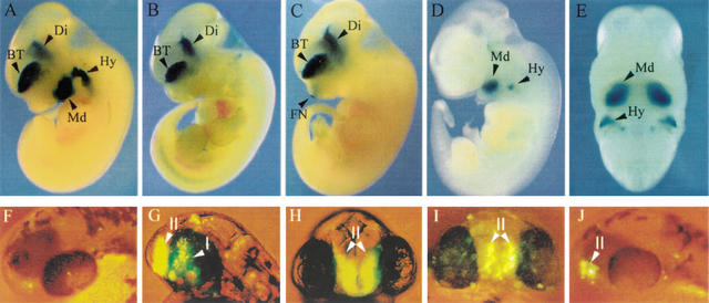
Enhancer activity of conserved Dlx intergenic sequences in transgenic mice (A–E) and zebrafish (F–J). (A) Mouse I56i, (B) mouse I56ii, and (C) mouse I12b each drive reporter gene expression to the telencephalon (BT) and diencephalon (Di) of transgenic mice, as shown here in mouse embryos. (D, E) Mouse I12a drives reporter gene expression to a subset of mesenchymal cells in the mandibular (Md) component of the first branchial arch and in the second branchial arch (Hy) of an E11.5 embryo. (A–D) are sagittal views and (E) is a frontal view of the embryo shown in (D). All embryos are at stage E11–12. FN, frontonasal prominence. (F) Head of 48 hpf primary transgenic zebrafish embryo, dorso-lateral view, injected with the control dlx6a-GFP reporter plasmid. Injection of this construct results in very few GFP-positive cells with no tissue specificity (n > 150). (G,H) Lateral and frontal views, respectively, of a 48-hpf zebrafish embryo from a transgenic line produced with a construct made with the dlx6a-GFP reporter plasmid that also contained a 1.4-kb dlx5a/dlx6a intergenic fragment containing I56i and I56 ii. I and II indicate the diencephalic and telencephalic domains of transgene expression, which also correspond to endogenous dlx expression patterns in the zebrafish forebrain. (I) Frontal view of a 48-hpf primary transgenic zebrafish embryo injected with a dlx6a-GFP that also contained a 4.0-kb mouse Dlx5/Dlx6 intergenic fragment that comprises I56i. The transgene is expressed predominantly in the telencephalic domain II. (J) Lateral view of a 48-hpf primary transgenic zebrafish embryo injected with a dlx6a-GFP that also contained a 2.8-kb mouse Dlx5/Dlx6 intergenic fragment that comprises both I56i and I56ii. GFP-positive cells are seen only in the telencephalic domain, II.

