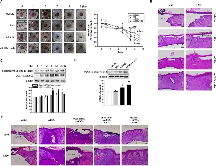
Figure 1.
Role of mESCs with shh on skin wound healing in vivo. Vehicle, shh, mESCs or mESCs with shh (mESCs + shh) were applied to the skin wound. (A) Representative appearance of wound tretated with vehicle, shh, mESCs, or mESCs + shh at days 0, 1, 5, 7, 8, 9 (left panel). Quantification of wound size relative to original wound size (day 0) is calculated (right panel). Data represents the mean ± SD (n = 6). *P < 0.05 versus day 0 of vehicle; #P < 0.05 versus day 0 of shh; &P < 0.05 versus day 0 of mESCs; +P < 0.05 versus day 0 of mESCs + shh. (B) Representative haematoxylin and eosin (H&E) staining of wound skin at day 9 after wounding (n = 6). Scale bar = 200 μm. (C) mESCs were treated with shh for various time (0–24 h), and the secreted VEGF into medium and the expression of VEGF in mESCs were detected by immunoblot. The lower panel depicts the mean ± SD of three independent experiments for each condition, as determined from densitometry relative to β-actin. *P < 0.05 versus 0 h of secreted VEGF into medium; #P < 0.05 versus 0 h of VEGF in mESCs. (D) The expression of VEGF was in wound skin at day 9 after wounding was determined by immunoblot. The lower panel depicts the mean ± SD of three independent experiments for each condition, as determined from densitometry relative to β-actin. *P < 0.05 versus vehicle; #P < 0.05 versus mESCs. (E) Vehicle, mESCs, VEGF siRNA-transfected mESCs [VEGF siRNA (20 nM) + mESCs], VEGF siRNA-transfected mESCs with shh (VEGF siRNA + mESCs + shh) or Nt siRNA-transfected mESCs with shh (Nt siRNA + mESCs + shh) were applied to the skin wound. Representative H&E staining of wound skin at day 9 after wounding. Scale bar = 100 μm.

