FIG 1.
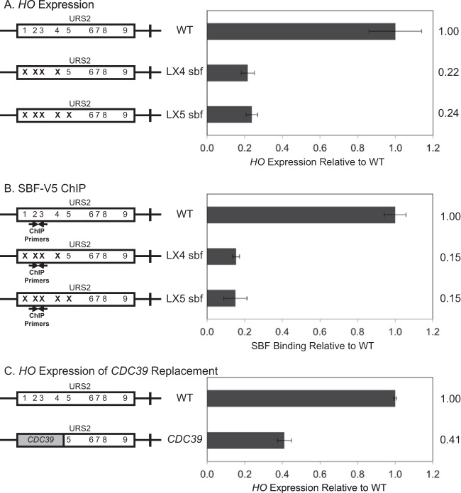
Downstream SBF sites cannot compensate for left-half site mutants. Diagrams on the left represent the URS2 portion of the HO promoter, and numbers within the rectangles indicate SBF site locations. Mutations in an SBF site are indicated by an X. CDC39 replacement is indicated by a gray block. (A) HO mRNA levels were measured for strains with the wild-type, LX4 sbf, and LX5 sbf versions of the HO promoter. (B) Binding of the Swi4-V5 subunit of SBF to the wild-type, LX4 sbf, and LX5 sbf versions of the HO promoter was determined by ChIP followed by qPCR. HO SBF ChIP enrichment was normalized to that of CLN1 and graphed relative to wild-type enrichment. Location of ChIP primers is indicated on the diagram. (C) HO mRNA levels were measured for strains with the wild-type HO promoter and CDC39 replacing URS2L.

