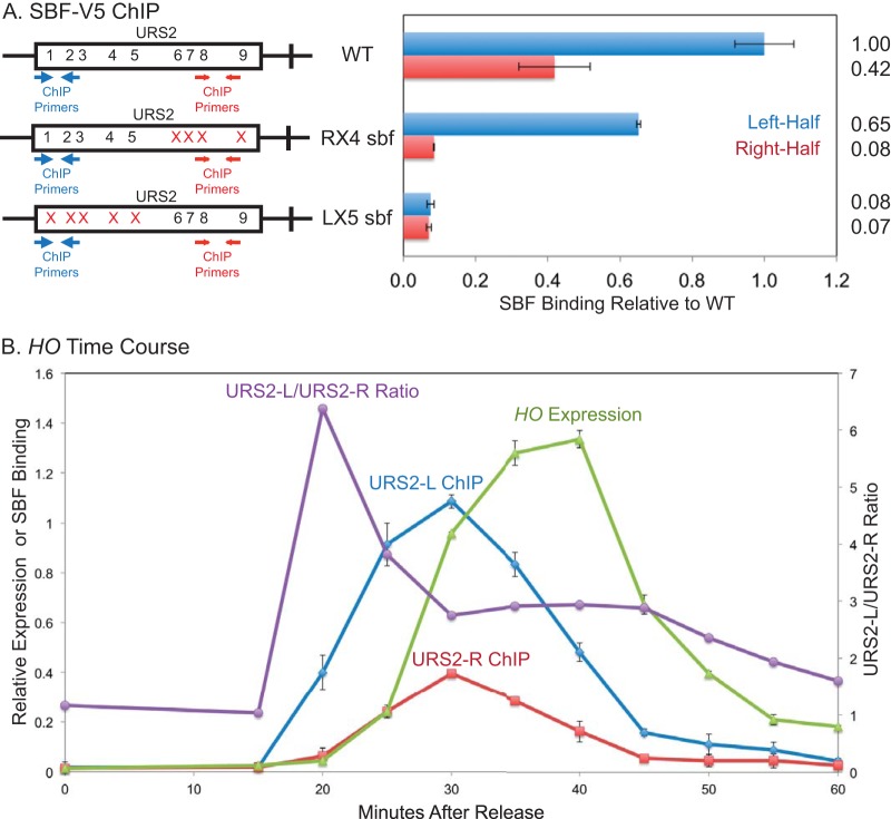
FIG 7.
Left-half SBF sites are required for SBF binding to the right half of URS2. (A) Diagrams on the left represent the URS2 portion of the HO promoter, and numbers within the rectangles indicate SBF site locations, with the location of ChIP primers indicated. Mutations in an SBF site are indicated by red X's. Binding of the Swi4-V5 subunit of SBF to the indicated versions of the HO promoter was determined by ChIP followed by qPCR. (B) Cells containing the GALp::CDC20 allele were synchronized by galactose withdrawal and readdition. The 0-min time point represents the G2/M arrest, before release. Cells were harvested at the indicated time points, and samples were processed for both RNA and chromatin. HO expression was measured by RT-qPCR, and binding of the Swi4-V5 subunit of SBF was determined by ChIP followed by qPCR. The ChIP values for URS2-L enrichment, URS2-R enrichment, and HO expression make use of the left y axis. The URS2-L/URS2-R ratio is plotted against the right y axis, and this represents the ratio of URS2-L enrichment divided by URS2-R enrichment. Error bars reflect the standard deviations from two independent ChIP experiments from the same set of synchronized cells.

