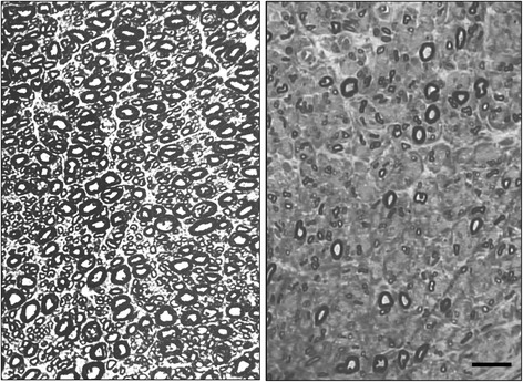
Figure 1.

Light microscopy. Photomicrographs of semithin sections. left figure: normal supraorbital nerve from patient 0. Note the densely packed myelinated fibres. right figure: supraorbital nerve from Patient 5 showing rarefied large-size myelinated fibres, whereas some small myelinated fibres are preserved. Bar: 20 μm.
