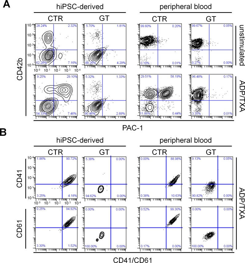
Figure 3. Flow cytometry of PAC-1 binding and integrin surface expression after platelet activation.
(A) Cells were stained with PAC-1 (x-axis) and anti-CD42b (y-axis) antibodies in the absence (top) or presence (bottom) of ADP/TXA2. (B) Flow cytometry of CD41 (GPIIb) and CD61 (GPIIIa) surface expression on hiPSC-derived and peripheral blood platelets in the presence of ADP/TXA2. After activation with ADP/TXA2, cells were stained with anti-CD42b, anti-CD41/CD61, anti-CD41 and anti-CD61 antibodies. The FSC/SSC log gate of peripheral platelets was applied and further gated for CD42b+ cells.

