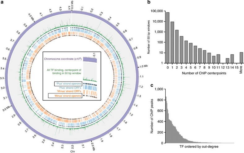
Figure 1. A global view of DNA binding.
(a) TF-binding sites identified by ChIP-seq plotted with Circos59. Sense (blue) and antisense (orange) CDS and operon boundaries illustrated with black edges. The 4.4-Mb H37Rv chromosome is divided into nonoverlapping 50-bp windows, and green spikes represent the total number of TF-binding events within each window. (b) Histogram of number of TF-binding events per 50-bp window. (c) Number of ChIP-binding events (out-degree) for each of the 156 DNA-binding proteins with at least one binding site.

