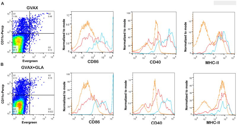
Figure 3.
Phenotypic analysis of APC activation markers in DLN on day 6. A) Representative analysis of mice vaccinated with GVAX alone (A) and GVAX+GLA (B). EverGreen+ CD11c+ cells (blue) show increased expression of activation markers CD86 and CD40 in mice vaccinated with both GVAX and GVAX+GLA compared to EverGreen-CD11c+ cells (red) and EverGreen-CD11c- cells (orange) in both groups. A population of EverGreen-CD11c+ cells also exhibit upregulated activation markers but not to the extent seen for EverGreen+CD11c+ cells. The expression level of MHC-II in EverGreen+CD11c+ cells (blue) in both groups was comparable to EverGreen- CD11c+ cells (red). The expression levels of these markers were not significantly different between the two vaccinated groups.

