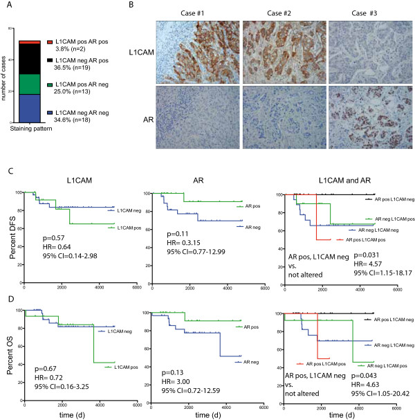
Figure 4.

Staining examples for L1CAM and AR breast cancer tissue sections. (A) Staining pattern, showing the number of cases of the TNBC Innsbruck cohort that were stained by IHC for L1CAM and AR. Details are summarized in Table 1 (n = 52). (B) IHC staining for L1CAM and AR on representative sections from the Innsbruck cohort. Kaplan Meier analysis of disease free survival (DFS) (C) and overall survival (OS) (D) of the Innsbruck cohort that were stained by IHC for L1CAM and AR.
