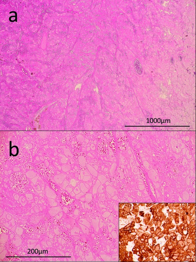
Fig. 3.

A: Hematoxilin-eosin (H&E) staining of the tumour tissue (×20). B: H&E staining (×100) and cytokeratin 7 staining (lower right) of the tumour tissue. The tumour was completely necrotic. Tumour cells with abundant cytoplasm and large nuclei formed nests. Tumour structure was intact.
