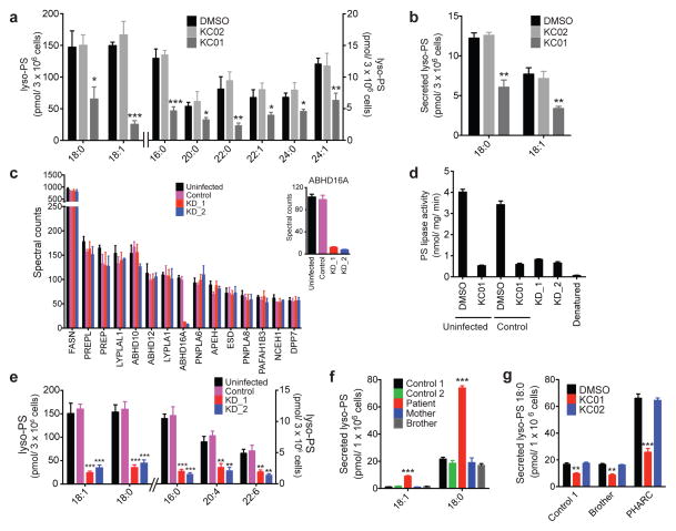
Figure 3. Disruption of ABHD16A reduces the lyso-PS content of human cells.
(a, b) Cellular (a) and secreted (b) concentrations of lyso-PS from COLO205 colon cancer cells treated in situ with inhibitors (KC01 or KC02, 1 μM) or DMSO for 4 h. See Supplementary Table 1 for complete list of quantified lipids. (c) Spectral count values of serine hydrolase activities of the membrane proteomes of the indicated cell models as measured by ABPP-MudPIT. Inset shows a blow-up of the ABHD16A spectral counts. Shown are the 15 most abundant serine hydrolase activities. See Supplementary Table 2 for the complete list of serine hydrolase activities. (d) PS lipase activities of membrane proteomes from the indicated K562 cell lines. Uninfected and control cell line proteomes were pre-treated with DMSO or KC01 (1 μM, 30 min, 37 °C) to establish PS lipase activities for baseline and ABHD16A-inhibited samples. The uninfected proteome was also heat-denatured prior to analysis as an additional control. (e) Cellular concentrations of lyso-PS from the indicated K562 cell lines. (f) Secreted concentrations of lyso-PS from the indicated lymphoblast cell lines (LCLs). (g) Concentrations of secreted C18:0 lyso-PS from the indicated LCLs treated with DMSO or inhibitor (KC01 or KC02; 1 μM; 4 h). Data represent mean values ± s. e. m for three-eight biological replicates. Student’s t-test: * p < 0.05; **p < 0.01; *** p < 0.0001 for experimental versus control groups.

