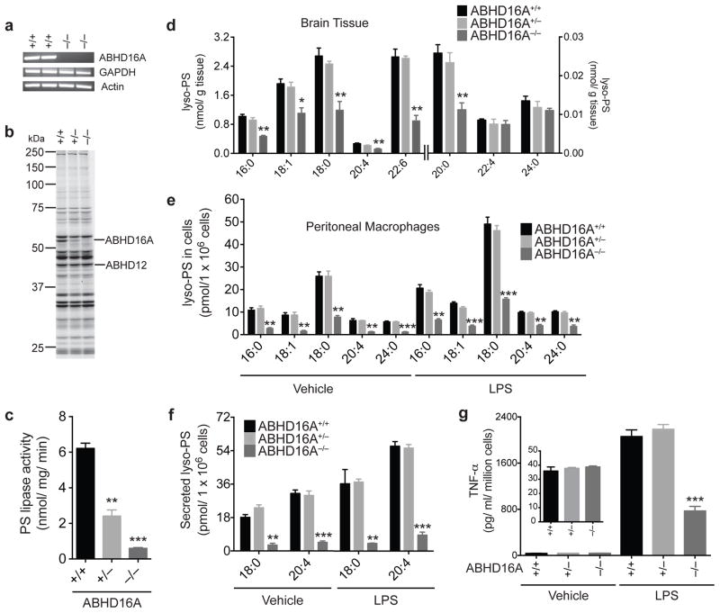
Figure 5. Generation and characterization of ABHD16A−/− mice.
(a, b) Confirmation of absence of ABHD16A mRNA and protein activity in brain tissue from ABHD16A−/− mice using RT-PCR (a) and ABPP (b) analyses, respectively. ABPP gel represents treatment of cerebellar membrane proteomes ABHD16A+/+, +/−, and −/− mice with FP-rhodamine (2 μM, 30 min, 37 °C). (c) PS lipase activity of brain membrane lysates from ABHD16A +/+, +/−, and −/− mice. Data represent mean values ± s. e. m. for three biological replicates. Student’s t-test: ** p < 0.0005, *** p < 0.0001 for ABHD16A−/− versus ABHD16A+/+ groups. (d) Concentrations of lyso-PS from brain tissue of ABHD16A+/+, +/−, and −/− mice. Data represent mean values ± s. e. m. for four biological replicates. Student’s t-test: * p < 0.05, ** p < 0.005, for ABHD16A−/− versus ABHD16A+/+ groups. (eg) Concentrations of cellular (e) and secreted (f) lyso-PS and TNF-α (g) from thioglycollate-elicited peritoneal macrophages derived from ABHD16A+/+, +/−, and −/− mice after treatment with vehicle (PBS) or LPS (5 μg/mL) for 7 h. Data represent mean values ± s. e. m. for four biological replicates Student’s t-test: ** p < 0.0005, *** p < 0.0001, ABHD16A−/− versus ABHD16A+/+ groups.

