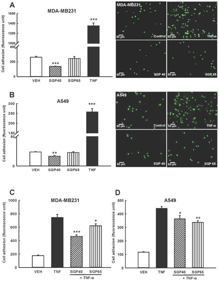
Fig. 2. Adhesion of tumor cells to human brain endothelial cells is attenuated by SGPs.
(A and B) Endothelial and calcein-labeled breast (A) or lung (B) tumor cells were exposed to SGP40, SGP65 (both normalized to 5 μM of Se), TNF-α (10 ng/mL, positive control), or vehicle (VEH) for 24 h. Representative fluorescence images from n=6 experiments are shown on right panels. Left panels show quantitative data from these experiments. (C and D) Endothelial and calcein-labeled tumor cells were exposed to SGP40, SGP65, or vehicle (VEH) as in (A and B). Then, TNF-α (10 ng/mL) or vehicle was added for an additional 20 h incubation. Values are mean ± SEM, n=6–8 wells per group. ***p<0.001, **p<0.01, *p<0.05 compared with vehicle (A and B) or TNF-α (C and D).

