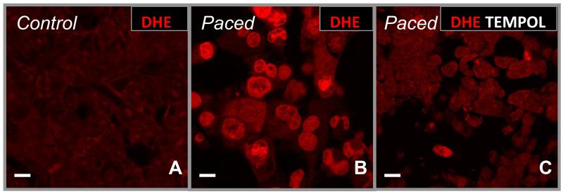
Figure 2. Production of superoxide (O2·−) with rapid stimulation.
After 6hr of rapid stimulation in culture, O2·− production by atrial HL-1 cells was detected by dihydroethidium (DHE; 10μM; B, indicated by the bright red color) that is not present in control, spontaneously-beating cells (A). Superoxide generation was essentially abolished under these conditions by co-incubation of cells with the cell-permeable superoxide dismutase mimetic tempol (C). Scale bars = 10 microns.

