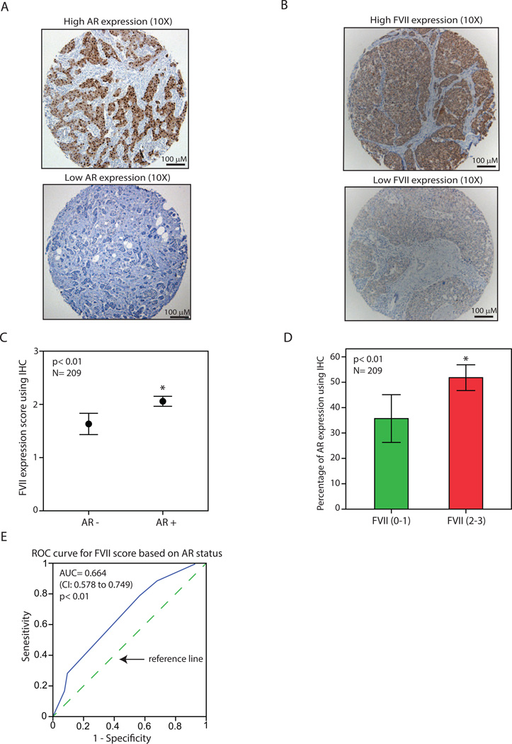
Figure 3.
Association of factor VII and AR protein expression in breast tumors using immunohistochemistry. (A) Immunohistochemistry (IHC) to demonstrate breast tumors with a high level (top panel) and a low level (bottom panel) of AR expression. Magnifications are at 10X. (B) IHC to demonstrate breast tumors with a high level (top panel) and a low level (bottom panel) of coagulation factor VII (FVII) expression. Magnifications are at 10X. (C) FVII expression scores using IHC for AR+ and AR- breast tumors. *, p< 0.01 is for AR+ vs. AR- groups. (D) Percentage of AR expression using IHC for breast tumors with a low FVII staining (scores: 0–1) and those with a high level of FVII (scores: 2–3). *, p< 0.01 is for FVII (2–3) vs. FVII (0–1). (E) Receiver operating characteristic (ROC) analysis to predict FVII-IHC scores based on the AR status in primary breast tumors. FVII scores ranged from 0–3 and AR status was 0 (< 10% AR staining) or 1 (≥ 10% AR staining). CI: confidence interval. Dashed line is a diagonal reference line. All Error Bars: ± 2SEM.

