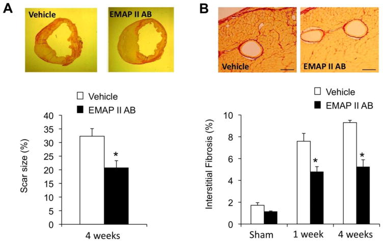
Figure 2.
EMAP II AB improved LV structure with reduced scar size and collagen deposition. (A) The scar size was decreased after EMAP II AB treatment for 4 weeks. Upper panels: Representative images of infarct rings after 4wk MI as shown by PSR staining; lower panel: quantitative data. (B) The interstitial fibrosis (%) in the adjacent area was less in the EMAP II AB group after both 1 and 4 week MI as shown by PSR staining. Upper panels: representative images after 1 wk MI; lower panel: quantitative data.

