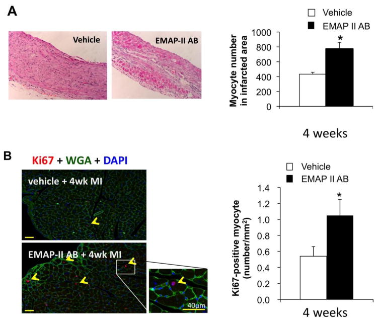
Figure 3.
EMAP II AB preserved myocytes and increased proliferating myocytes after chronic MI. (A) More preserved myocytes were seen in EMAP II AB as shown with H&E staining of infarct area after 4 week MI. Left panels: H&E staining images (original magnification, ×10); right panel: quantitative data. (B) Proliferating myocytes were increased with EMAP II AB as shown by Ki67-WGA-DAPI triple staining (left panel) and quantification (right panel). *: p<0.05 compared with vehicle. n=6 for each group.

