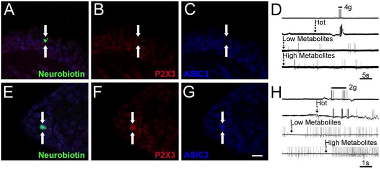
Figure 6. Examples of intracellularly stained and physiologically characterized muscle sensory neurons recovered from the ex vivo recording preparation after ischemic muscle injury (prolonged ischemia or transient ischemia with reperfusion).

A muscle afferent (arrows) intracellularly filled with neurobiotin (A, green) was recovered from an ex vivo preparation in a mouse after ischemic muscle injury was found to be responsive to mechanical stimuli, hot saline and both low and high metabolite mixtures (D) was found to be immunonegative (arrows) for both P2X3 (B, red) and ASIC3 (C, blue). Another neurobiotin filled (E, green) muscle afferent was found to be responsive to mechanical stimuli, hot saline and both low and high metabolites (H) was found to coexpress (arrows) both P2X3 (F, red) and ASIC3 (G, blue). Some spontaneous activity was present in the latter cell prior to metabolite stimulation. Scale bar for all images, 40μm. Dashed arrows in D and H indicate onset of stimulus in the muscles.
