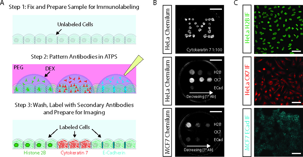
Figure 1. Multiplexed immunostaining of cell monolayers.

(A) Aqueous two-phase system-mediated multiplexed immunostaining uses ATPSs composed of PEG and dextran to micropattern primary antibody solutions on the surface of the sample. Apart from the primary antibody incubation step, we follow standard immunostaining procedures. (B) Chemiluminescence detection of cytokeratin 7 (CK7), histone 2B (H2B) and E-cadherin (ECad) in antibody-micropatterned HeLa and MCF7 cell monolayers. The top image shows a HeLa monolayer immunostained in a “Michigan M” pattern using 23 dextran droplets containing 1:100 anti-CK7 antibody. The middle and bottom images show cell type-specific staining for H2B (control), CK7 and Ecad in HeLa cells and MCF7 cells, respectively. From left to right the antibody dilution were 1:100, 1:400 and 1:800 for the anti-H2B antibody, 1:100, 1:600 and 1:1000 for the anti-CK7 antibody and 1:100, 1:600 and 1:1500 for the anti-ECad antibody. The spacing between the primary antibody spots can be estimated from the scale bars, which are ~10 mm. (C) Immunofluorescence detection of antigens at dextran /antibody micropatterned spots for H2B (top, 1:1000 dilution), CK7 (middle, 1:100 dilution) and ECad (bottom, 1:400 dilution). Scale bars are ~50 µm.
