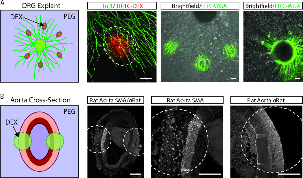
Figure 2. Multiplexed immunostaining of tissue explants and histological sections.

(A) DRG explants from chick embryos were selected to demonstrate the potential of this technique for multiplexed immunostaining of complex samples. The micropatterning system was initially tested by localizing TRITC- dextran -containing dextran microdroplets to the axons of Tuj1-labeled DRG explants (left-two images). We next tested the ability to micropattern biochemical stains, such as FITC-wheat germ agglutinin (FITC-WGA). FITC-WGA staining could be used to selectively label both the axons and ganglia of the DRG explants (right-two images). Scale bars are ~100 µm. (B) Multiplexed immunostaining of histological section was demonstrated on paraffinized cross-sections of rat abdominal aortas using anti-α-smooth muscle actin (SMA) and anti-rat antibodies. Scale bars are ~200 µm.
