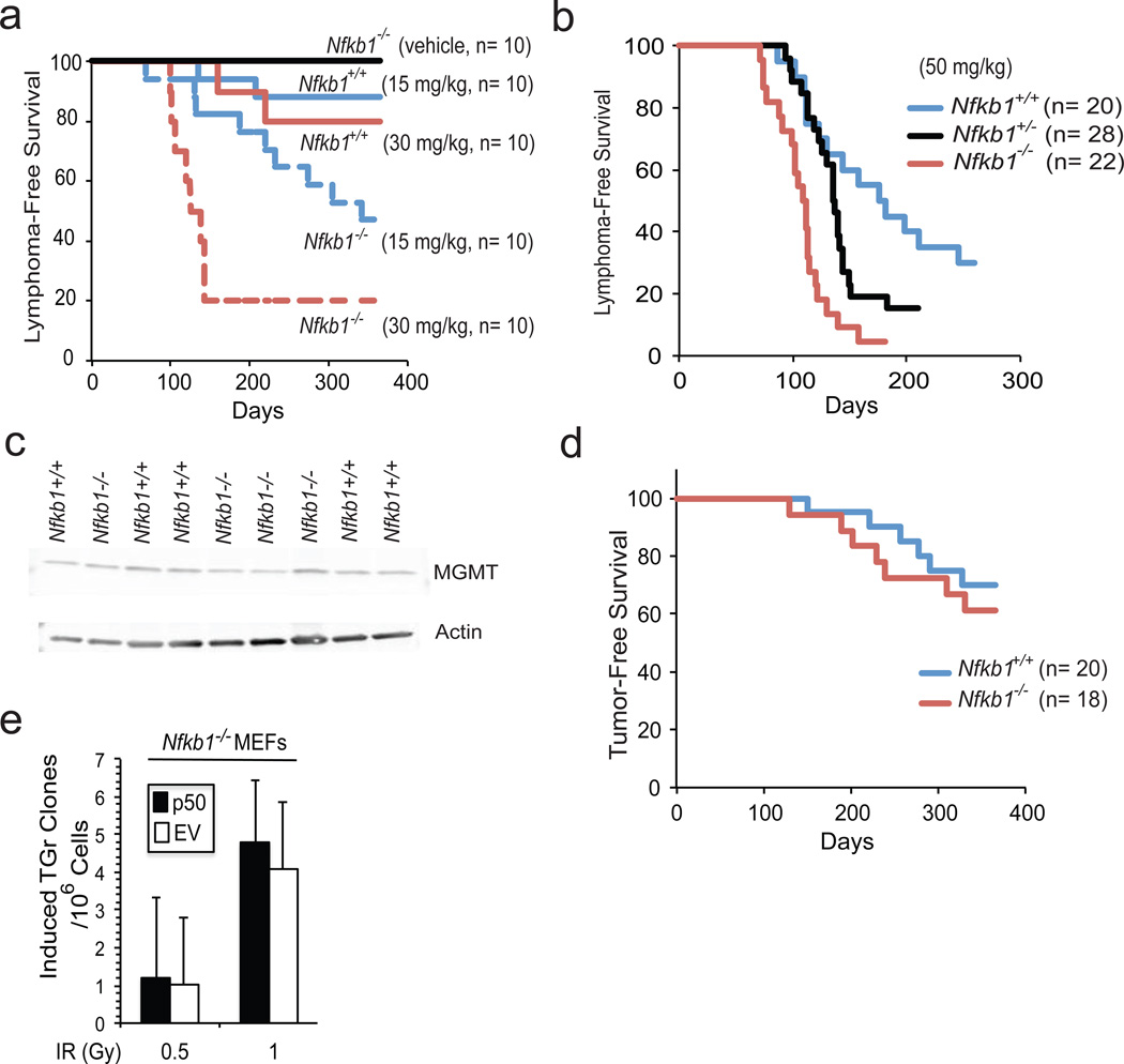
Figure 2.
Nfkb1 prevents alkylator-induced tumor formation. (a, b and d) Kaplan-Meier survival curves in mice following a single i.p. injection of MNU. (a) B6/129 Nfkb1+/+ and Nfkb1−/− animals treated with two concentrations of MNU. Statistical significance: P < 0.002 at 30 mg/ kg MNU and P < 0.02 at 15 mg/kg. (b) C57BL/6 mice treated with 50 mg/kg MNU. Statistical significance: P < 0.0001: Nfkb1+/+ vs. Nfkb1−/−; P = 0.05: Nfkb1+/+ vs. Nfkb1+/−; and P = 0.002: Nfkb1+/− vs. Nfkb1−/−. (c) Western blot with anti-MGMT antibody in thymus extract of untreated C57BL/6 Nfkb1+/+ and Nfkb1−/− mice. (d) C57BL/6 Nfkb1+/+ and Nfkb1−/− animals treated with whole body IR. Statistical significance: P > 0.5. (e) Hprt assay in Nfkb1−/− MEFs stably expressing p50 or empty vector (EV) following treatment with IR. Animal experiments were performed in accordance with the guidelines of the Institutional Animal Care and Use Committee of the University of Chicago. Nfkb1−/− mouse strains used include B6;129P-Nfkb1tm1Bal/J and B6.Cg-Nfkb1tm1Bal/J that have been backcrossed for 12 generations with C57BL/6J mice (Jackson Laboratories, Bar Harbor, Maine, USA). The respective Nfkb1+/+ controls include: B6;129PF1/J and C57BL/6J. For tumorigenesis in the B6;129P strain, male mice were used and for tumorigenesis in C57BL/6J mice, equal ratios of male to female animals of all genotypes were used. Animals were housed in a pathogen-free, biosafety level II facility. MNU was dissolved in DMSO and diluted in PBS within 10 min of injection. The final preparation was delivered in a total volume of 500 µl per 25 g mouse. Six-week old mice were given a single i.p. injection of MNU or vehicle. For radiation carcinogenesis, equal ratios of male to female animals were treated with four weekly doses of 1 Gy total body irradiation using a Phillips RT250 x-ray generator. Animals were observed at least 2 times per week and sacrificed when they exhibited signs of cachexia, hunched position, respiratory distress or a visible mass. Following euthanasia, autopsy was performed. For evaluation of thymic MGMT expression, 5– 8 week old male C57BL/6 mice were sacrificed and the thymus harvested. Immunoblot was performed on cell lysates with anti-MGMT antibody (sc-8828, Santa Cruz Biotechnology, Dallas, Texas, USA). For histological analysis, tumors were removed and either snap frozen, disaggregated for FACS analysis or immersed in paraformaldyhyde prior to sectioning. Statistical analysis was performed using one-sided Mantel-Cox Log rank analysis.

