Figure 3.

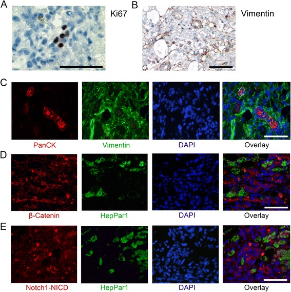
Proliferation and mesenchymal characteristics on HPCs and relation of differentiation with β-catenin and Notch1/NICD signalling. Immunohistochemical staining for Ki67 shows positive cells in the ductular reaction during LDH (A). Immunohistochemical staining for vimentin suggests positive ductular reactions (B) and an example of vimentin and PanCK double staining (C) shows clear co-localisation on HPCs in LDH. Double immunofluorescence against HepPar1 and β-catenin (D) or Notch1/Notch intracellular domain (Notch1/NICD; (E) in LDH shows polarisation of the ductular reaction: clear cytoplasmic staining of β-catenin or Notch/NICD is present in non-differentiated cells of the ductular reaction, and only membranous staining is present in differentiating and fully differentiated, HepPar1 positive, hepatocytes. Size bars indicate 50 μm.
