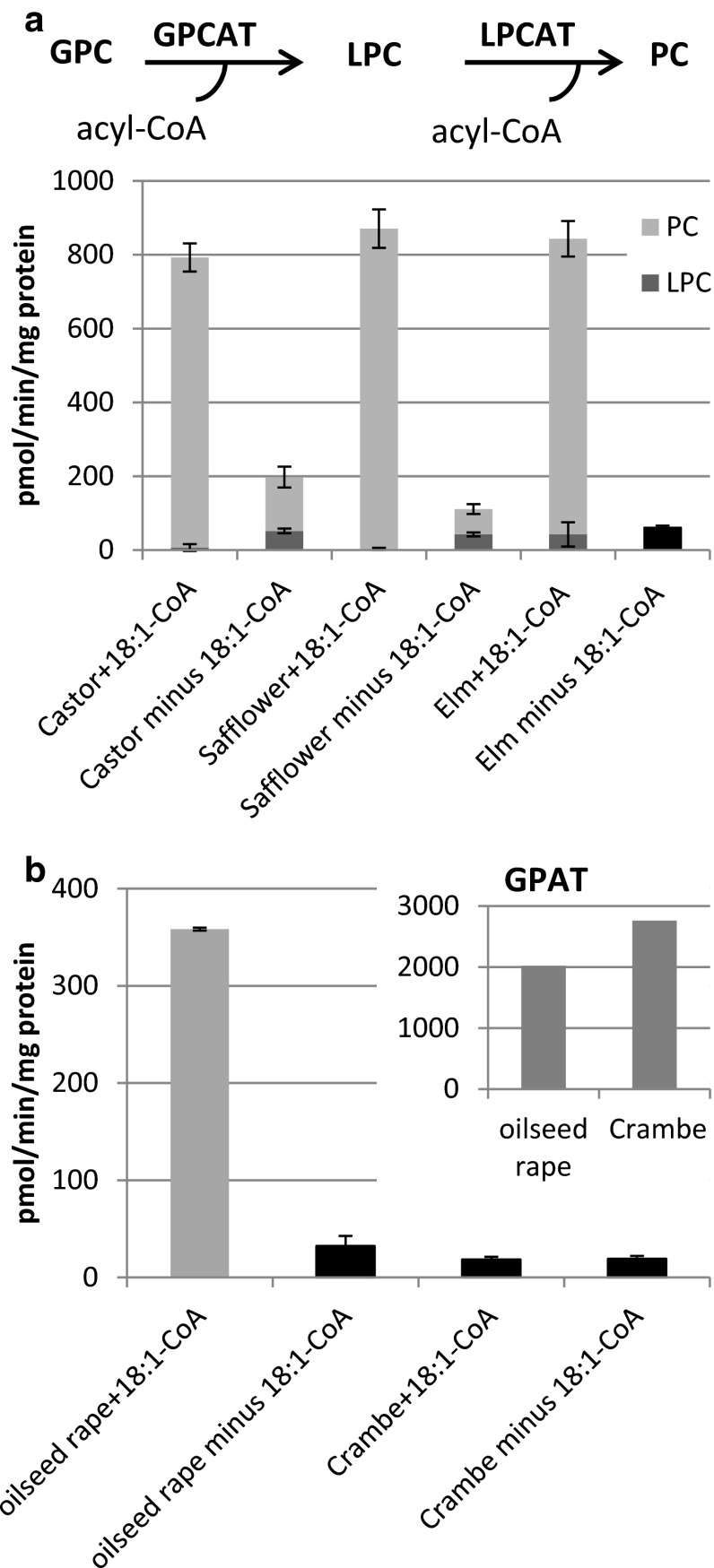
Fig. 1.
GPCAT activities in microsomal preparations from various developing oil seeds. The dark grey part of the bar represents the amount of radioactive LPC formed and the light grey part the amount of radioactive PC formed. Due to too low activity in membranes from Crambe and elm seed and rape in the absence of added acyl- CoA it was not possible to get accurate readings of the proportions of radioactivity between PC and LPC and the black bars represent the total amount of radioactivity incorporated into the chloroform fraction (the [14C]glycerophosphocholine substrate resides exclusively in methanol/water phase after extraction). a GPCAT activity with and without addition of 18:1-CoA. The combined enzyme reactions of LPCAT and GPCAT in the microsomal preparations are depicted at the top of the figure. b GPCAT and GPAT (inserted diagram) activities in microsomal preparations from developing seeds of oil seed rape (Brassica napus) and Crambe abyssinica in the presence and absence of 18:1-CoA. Microsomal membranes were re-suspended in buffer containing 0.1 % BSA and re-pelleted before used in assays. Microsomes corresponding to 40 μg were incubated with 25 nmol of [14C]glycerophosphocholine (specific activity 4,750 dpm/nmol) or 10 nmol of [14C]glycerol 3-phosphate (specific activity 6,100 dpm/nmol) for 20 min at 30 °C. All GPCAT assays were done in triplicate and values are shown ± SD

