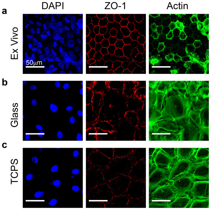
Figure 1. CE cells lose differentiated morphology when cultured on glass or tissue culture polystyrene.
(a) Example of ex vivo, intact endothelium from the bovine cornea where cells exhibit a polygonal, mostly hexagonal shape and small size with the tight junction protein ZO-1 (red) present at cell borders and F-actin (green) located cortically. (b) CE cells cultured for one passage on a rigid glass substrate have reduced ZO-1 localization at the cell-cell borders and F-actin fibers present cortically as well as throughout the cell body. (c) CE cells cultured on TCPS for one passage appear similar to CE cells on glass, showing a loss of phenotypic shape and ZO-1 and F-actin localization relative to the ex vivo tissue. Cells are stained for nuclei (blue), tight junction protein ZO-1 (red) and F-actin (green) and scale bars are 50 μm.

