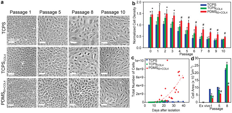
Figure 3. CE cells cultured on the biomimetic PDMS50+COL4 substrate maintained a polygonal morphology, higher cell density, increased proliferation rate and smaller cell size.
(a) Representative phase contrast images showing the morphology of CE cells cultured on the three different substrates at four different passages. At P5, CE cells cultured on TCPSCOL4 and TCPS exhibited elongated, irregular cell morphology. By P8 the cells were enlarged and polarized with no resemblance to a hexagonal morphology. In contrast, CE cells cultured on PDMS50+COL4 maintained a hexagonal like morphology up to P8. (b) Normalized cell density for CE cells cultured on TCPS (n = 4), TCPSCOL4 (n = 5) and PDMS50+COL4 (n = 5) (mean ± s.d.). At all passages the density of CE cells on the PDMS50+COL4 was significantly greater than on the TCPS and than on the TCPSCOL4 from P4 to P10. (c) Number of CE cells on the different substrates as a function of time, where each data point represents cell number prior to passaging (up to P5 for TCPS and TCPSCOL4 and P8 for PDMS50+COL4, when CE morphology became non-polygonal). There was a >3000-fold increase in the total cell number on PDMS50+COL4 compared to approximately a 140-fold increase on TCPS and TCPSCOL4 (dashed lines are to guide the eye). (d) Cell area in the ex vivo endothelium and on TCPS, TCPSCOL4 and PDMS50+COL4 at P1, P5 and P8 (mean ± s.e.m.). (*) indicating a statistically significant difference (P < 0.05) between PDMS50+COL4 and TCPS and (#) indicating a statistically significant difference (P < 0.05) between PDMS50+COL4 and both TCPS and TCPSCOL4.

